Methods
Overview
- histology, immunohistochemistry, and tracer backfills
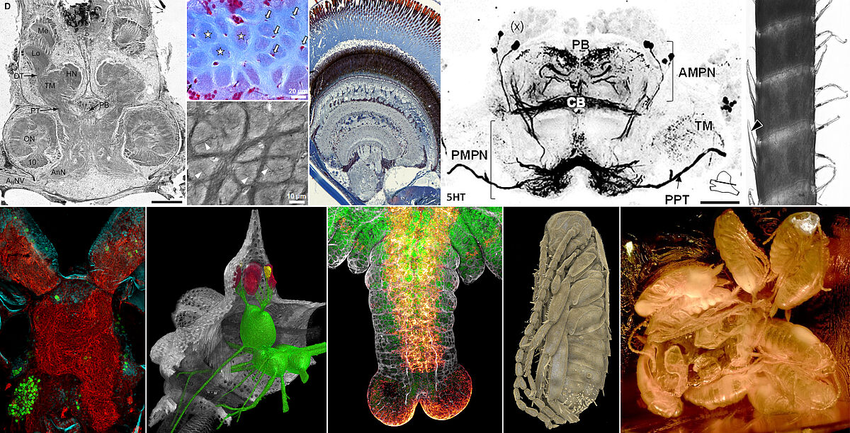
- imaging techniques and 3D reconstruction
- electrophysiology
- behavioral experiments in the field and in the lab, bioessays
Sectioning devices
- vibratome Zeiss Hyrax V-50
- rotary microtome Leica RM 2145
- rotary microtome Microm HM 360
- sliding microtome Leica SM2000 R

Optical equipment
(see also Imaging Center)
- macro- and microphotographic setup
- fluorescence microscope with co-observation module Nikon SMZ800N
- epifluorescence microscope Nikon Eclipse 90i
- light microscope Zeiss Axiophot
- scanning electron microscope Zeiss EVO LS10
- transmission electron microscope Jeol JEM 1011
- confocal laser scanning microscope Leica SP5 II
- X-ray micro-computed tomograph Zeiss Xradia XCT-200
Visualisation software and 3D printing
- Bitplane Imaris
- FEI Amira
- Ultimaker 2 (3D Printer)
