Theses
Current topics for B.Sc. and M.Sc. theses
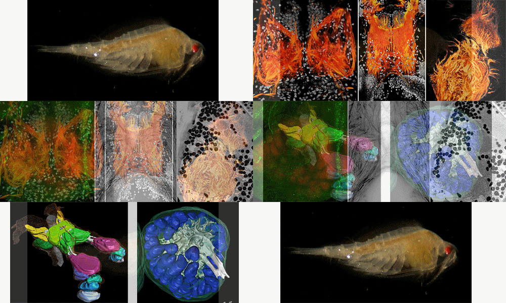
Methods: Macrophotography; immunhistochemistry against cytoskeletal proteins, neuroactive substances (e.g. neurotransmitters and -modulators) and synaptic proteins; neuronal backfills; computer-aided 3D reconstruction; histological sectioning; fluorescence microscopy; confocal laser-scanning microscopy; computer-based X-ray tomography; scanning electron microscopy (SEM).
If you prefer to, you may propose your own project for your B.Sc. or M.Sc thesis. Especially if relying on some of the methods listed above, we can discuss the feasibilty of such proposals at any time. In addition, you can choose research topics advertised by our group. The exact scope and focus of your personal project will then be adapted depending on the time available (in accordance with B.Sc.- or M.Sc. theses guidelines).
Contact:
- Prof. Dr. Steffen Harzsch
- Dr. Marie K. Hörnig
- Dr. Sebastian Büsse
Graduated theses of the department
- (Lena Hoffmann, B. Sc. - "Using immunohistochemistry against allatostatin to describe sex differences in the brain and the olfactory pathway of Neomysis integer")
- Johanna Blatt, B. Sc. - "Using immunohistochemistry against RFamide to describe sex differences in the brain and the olfactory pathway of Neomysis integer"
- Leon Rostock, M. Sc.
- Kerstin Lipinske, M. Sc. - "Der Eurasische Fischotter (Lutra lutra) an der Barthe (MV): Nahrungsanalyse und Interpretation trophischer Zusammenhänge im Verlauf der Jahreszeiten"
- Natalie Kirstein, B. Sc. - "Ontogenetic aspects of extinct predatory orthopterans – Gryllobencain patrickmuelleri as example"
- Frederice Hilgendorf, M. Sc. - "Neuroplasticity in the brain of the marbled crayfish Procambarus virginalis (Lyko, 2017): Impact of different rearing regimes on structure and allometric growth of the hemiellipsoid body"
- Linus Hübner, B. Sc. - "Etablierung eines Immunfluoreszenz-Nachweises der Cannabinoid-Rezeptoren CB1 und CB2 für den Maus-Skelettmuskel"
- Marco Schade, Promotion Dr. rer. nat. - "Fancy skulls and simple minds: (neuro)anatomical implications for palaeobiology of non-avian dinosaurs"
- Carina Paetzel, M. Sc.
- Isabel Grajewski, B. Sc. - "Vergleich konventioneller Antikörper mit Einzeldomänenantikörpern (Nanobodies) im Nervensystem von Procambarus virginalis anhand bildgebender Parameter in der Immunhistochemie"
- Sophie Raspe, B.Sc. - "Characterization of SIFamide-stainings in the brain of Parhyale hawaiensis"
- Sara Zielke, M.Sc - "Habitat choice and food preference of Rhithropanopeus harrisii in Vitter Bodden, in the southern Baltic Sea"
- Sarah Zok, M.Sc - "Evolution der Ontogenese bei Blattodea - Außenmorphologischer Vergleich rezenter und fossiler Vertreter"
- Zoran Šargač, Promotion Dr. rer. nat. - "Global ocean change: responses of crab larvae to abiotic drivers"
- Pia Steingrübner, M. Sc. - "Effect of temperature fluctuations on larval development and survival of the Asian shore crab Hemigrapsus sanguines"
- Hanna M. Dedenbach, M.Sc - "Modellierung der Vorkommen von Schweinswalen in der Ostsee und angrenzenden Gewässern anhand von Zufallssichtungen durch Wassersportler"
- Janush A. Axmann, B.Sc - "Seasonal reproduction of female rays from the Chilean round ray (Urotrygon chilensis) and Haller's round ray (Urobatis halleri) in the northern Pacific of Costa Rica"
- Christin Wittfoth, Promotion Dr. rer. nat. - "Comparative aspects of adult neurogenesis and the central olfactory pathway in malacostracan crustaceans"
- Henning Hoffmann, B.Sc - "Mageninhaltsanalysen der Kegelrobbe (Halichoerus grypus) in der deutschen Ostsee"
- Marie K. Hörnig, Promotion Dr. rer. nat. - "Rekonstruktionen zur Evolution von Fortpflanzungsstrategien und Raubverhalten geflügelter Insekten: Was Fossilien über komplexe Verhaltensweisen verraten"
- Franziska Spitzner, Promotion Dr. rer. nat. - "Effects of environmental stress on early life history stages of the European shore crab Carcinus maenas Linnaeus 1758 (Brachyura: Portunidae)"
- Rebecca Meth, M.Sc. - "Die postembryonale Entwicklung des deutocerebralen proliferativen Systems bei Carcinus maenas (Brachyura, Decapoda)"
- Alexander Bracke, Promotion Dr. rer. nat. - "Untersuchung zur Auswirkung von Adipositas auf hippocampale Morphometrie und neuronale Plastizität sowie Verhalten in einem Leptin-defizienten Mausmodell" (in Kooperation mit der Universitätsmedizin Greifswald)
- Dr. Andy Sombke, Habilitation Dr. habil. - "Beiträge zur Morphologie und Evolution der Arthropoda mit einem Fokus auf das periphere und zentrale Nervensystem"
- Johannes Kaelke, M.Sc. - "Stable isotope analysis of otolith growth zones in Baltic cod (in Kooperation mit dem Thünen-Institut für Ostseefischerei Rostock)"
- Emanuel S. Nischick, M.Sc. - "Vergleichende Untersuchungen der Anatomie der Zoea V von Hemigrapsus sanguineus (Decapoda: Brachyura: Grapsoidea) unter abiotischem Stress"
- Jan D. Roy, M.Sc. - "Die Terminalbeine der Geophilomorpha - Analyse transformierter Arthropodien"
- Dipl.-Biol. Carlos A. Martínez-Muñoz, M.Sc. - "Preliminary conservation assessment of Cuban giant centipedes (Chilopoda: Scolopendromorpha)"
- Ann-Christin Rath (geb. Richter), M.Sc. - "Erfassen der Umwelt- die Morphologische Vielfalt der ersten und zweiten Antennenanhänge innerhalb der Malacostraca" (in Kooperation mit dem Meereskundemuseum Stralsund)
- Janine Voss. M.Sc. - "Vergleichende Untersuchungen zum Echoortungsverhalten von Schweinswalen (Phocoena phocoena) an repräsentativen Standorten der deutschen Nord- und Ostsee" (in Kooperation mit dem Meereskundemuseum Stralsund)
- Valerie Kreifelts, M.Sc. - "Neuroanatomy of Caprella mutica (Crustacea, Amphipoda, Caprellidae)"
- Christina Krüger, M.Sc. - "Der Einfluss von abiotischem Stress auf die larvale Muskulatur in Carcinus maenas (Brachyura, Decapoda)"
- Vanessa Schendel, M.Sc. - "Das Bauchmark von Lithobius forficatus (Chilopoda): Eine multimethodische Analyse mittels Histologie, Mikro-CT, Immunhistochemie und Backfilling"
- Dr. rer. nat. Christian W. Hädicke - "Walking on six legs: Einblicke in die Sinneswelt des Urhexapodens und die Evolution des Nervensystems der Mandibulata"
- Lukas Krautschick, B. Sc. - "Altersabhängige Volumenänderungen im Gehirn der Hornisse Vespa crabro"
- Dr. rer. nat. Matthes Kenning - "Evolution des olfaktorischen Systems der Isopoda: Einblicke aus Neuroanatomie und Ethologie von Saduria entomon (Valvifera) Linnaeus, 1758"
- Dr. rer. nat. Jakob Krieger - "Neurethologische Untersuchungen am größten terrestrischen Gliederfüßer, dem Palmendieb Birgus latro (Crustacea, Anomura) auf der Weihnachtsinsel (Indischer Ozean)"
- Emanuel S. Nischik, B. Sc. - "Morphometrische Analysen am Gehirn des Marmorkrebses Procambarus fallax f. virginalis: Vergleich verschiedener bildgebender Verfahren in der Neuroanatomie"
- Rebecca Meth, B. Sc. - "Die Anatomie des Gehirns von Penaeus vannamei"
- Caroline Viertel, M.Sc. - "Das Gehirn von Lithobius forficatus: Eine immunhistochemische Studie"
- Steinhoff Philip, B.Sc. - "Anatomy and neuroplasticity of the brain in the spider Marpissa mucosa (Salticidae, Arachnida)"
- Dipl.-Hum.Biol. Marie K. Hörnig
- Dipl.-Hum.Biol. Josephine Brennecke - "Immunohistochemistry in the olfactory lobe of Scutigera coleoptrata (Linnaeus, 1758) (Chilopoda)"
- Dipl.-Biol. Tina Kirchhoff
Gehirnarchitektur ausgewählter Taxa der terrestrischen Brachyura (Crustacea, Decapoda)
Diploma thesis by Dipl.-Biol. Philipp Braun
Although most members of the crustaceans can be found in aquatic habitats, several representatives are terrestrial. At least six major crustacean lineages independently evolved a semi- to fully terrestrial lifestyle (Ostracoda, Isopoda, Amphipoda, Brachyura, Anomura and Astacida). In my diploma thesis, I focus on the brain architecture of different taxa of terrestrial brachuyrans (Gecarcoidea natalis, Geosesarma tiomanicum, Cardisoma armatum and Uca tangeri). Therefore, I use different techniques like immunohistochemical labeling of brains and eyestalks using classical light-microscopy as well as confocal laser-scanning microscopy (cLSM). In addition, three dimensional-reconstructions of primary and secondary chemosensory processing areas is used for morphometric analyses between the selected species. Recent studies have shown that neuroanatomy reflects different terrestrial adaptations e.g. in anomurans and brachyurans. Thus, the aim of this comparative study is to understand more about the neuronal system and its terrestrial adaptation in different brachyuran species.
Rekonstruktion der multizellulären Ventraldrüsen von Spio filicornis
Bachelor thesis by Anton Rößger (extern, Uni Rostock)
Pigment-dispersing Hormone (PDH) Immunreaktivität im embryonalen Gehirn des Marmorkrebses
Bachelor thesis by Caroline Viertel, B.Sc.
Aspects of brain morphogenesis in the parthenogenetic marbled crayfish (Procambarus fallax virginalis, Hagen 1870)
Diploma thesis by Dipl.-Biol. Elisabeth Zieger
In this diploma thesis, the ontogeny of several neurotransmitter systems was documented in the embryonic brain of the Marbled Crayfish, using immunohistochemical methods and antisera against synapsin, serotonin, allatostatin, GABA, FMRFamide and histamine. For a direct comparison, embryos of the Migratory Locust were processed in the same way. Furthermore, the results were compared to literature data, to gain insight into common neurogenic features of insects and crustaceans.
In general, the anterior portion of the nervous system develops faster than the posterior portion. For each of the tested neurotransmitters, the immunoreactive structures that arise first bear remarkable morphological resemblance with each other. In both species, an initial step appears to be the establishment of a connection between the brain hemispheres. Fibres crossing the median protocerebrum of the Migratory Locust and the Marbled Crayfish exhibit immunoreactivity to the studied neurotransmitters particularly early. They are followed by neurites interconnecting the protocerebrum, the deuto- and tritocerebrum and the ventral nerve cord. The complex innervation patterns of specific neuropils, such as the central body, the accessory lobe, the olfactory lobe and the antennal lobe, on the other hand, emerge considerably later. Shortly before hatching, all the major neuronal pathways can already be seen, that are also present in adult locusts or crayfish. This suggests that all neurotransmitter systems investigated here are mainly operative by the time the embryos emerge. The pattern of immunoreactivity to a specific neurotransmitter was observed to always be quite similar between the Marbled Crayfish and the Migratory Locust. Accordingly, homology was proposed for certain neurons.
Brain architecture of Daphnia magna Straus, 1820 (Crustacea, Branchiopoda): Immunhistochemistry and 3D reconstruction
Diploma thesis by Dipl.-Biol. Timm Kress
In the present report, a detailed 3-dimensional reconstruction of the brain of Daphnia magna is presented. Most of the protocerebral neuropils as well as one deutocerebral neuropil could be reconstructed based on a series of osmium-ethylgallate stained semithin sections. Furthermore, immunolocalization experiments were carried out with antisera against synaptic proteins (synapsins), tyronisated tubulin, the neuropeptides FMRFamide, FLRFamide, corazonin and the biogenic amines histamin, and serotonin. The arrangement of immunolabeled neurons, patterns tracts and commissures in all neuropils of the protocerebrum and the deutocerebrum were described. These patterns were compared to data in other analyzed Branchiopoda.
About microevolutionary adaptations of ommatidia in the compound eyes of some Palaemon shrimp species (Caridea, Decapoda, Crustacea) on the fine structural level
Diploma thesis by Dipl.-Biol. Daniel Hamm (extern, Uni Regensburg)
Decapod shrimps of the genus Palaemon inhabit shallow areas of rocky shores across European coasts. Seven species are known to occur in Europe. Even though Palaemon species are found in high frequencies in the upper infralittoral, they display quite a ecological separation. On shallow rocky boulder fields in Ibiza, the study area of this diploma project, there are three different Palaemon species. Each of them populates a unique ecological niche of varying light exposition. Palaemon elegans is a diurnal rockpool species that seems to tolerate high light intensities. Palaemon xiphias inhabits the dim light world of Posidonia oceanica - seagrass beds. The troglodytic Palaemon serratus however avoids sunlight and can only be seen at night while scavenging in the upper infralittoral. The aim of this diploma project is to find out more about short-termed evolutionary effects of habitat segregation that might have affected visual capacities of those three Palaemon shrimps. Thereby, focus is not only on the microevolutionary transformation of the ommatidial architecture in their compound eyes, also the efficiency of photoperiodic adaptations within the ommatidia is of greater interest. As main methods to conduct this work we use light and transmission electron microscopy (TEM).
- Dr. rer. nat. Stefan Fischer
- Lisa Spotowitz, B.Sc.
- Lisa Thiele, B.Sc.
Comparative morphology of the brain of selected Diplopoda (Myriapoda) with focus on the central olfactory pathway
Diploma thesis by Dipl.-Biol. Florian Seefluth
Besides hexapods and crustaceans, the myriapods are the third arthropod taxon within the Mandibulata. The position of the Myriapoda within the Euarthropoda is still in debate. In the traditional system of Heymons (1901), the Myriapoda were the sistergroup to the Hexapoda. Recently, this hypothesis was challenged by two controversial alternatives: the Myriochelata concept (Chelicerata and Myriapoda) and the Tetraconata concept (Hexapoda and Crustacea). Following the latter concept, the Myriapoda represent the sistergroup to the Tetraconata (Regier et al. 2010). In the last decade, comparative neuroanatomical studies have provided a fresh view on arthropod relationships so that an analysis of Myriapoda brain architecture may contribute to our understanding of arthropod brain evolution (“Neurophylogeny” Harzsch 2006). However, comparative studies on the myriapod nervous system are few in number. Besides recent anatomical studies on the Chilopoda (Sombke et al. 2010), brain architecture in this taxon has not been studied in any detail. In order to gain more insights into the brain morphology of myriapods, we focus on the central nervous system of diplopods using immunohistochemical methods combined with fluorescence and laser scanning microscopy, scanning electron microscopy, anterograd backfilling, serial-thin section and 3D-reconstructions. Thus, several questions can be asked: how are the olfactory neuropils organized and does the brain architecture corresponds to other mandibulate taxa? In a neurophylogenetic approach, our diplopod data will be used for reconstructing arthropod phylogeny and for understanding the evolution of arthropod olfactory systems.
Gehirnarchitektur von Nebalia sp. (Leach, 1814) – Morphologie des zentralen Nervensystems eines basalen Malakostraken
Diplomarbeit von Dipl.-Biol. Matthes Kenning
Die Leptostraca (Packard, 1879) sind ubiquitär verbreite, doch obligat marine Crustacea, die von der Arktis bis in die Antarktis, in Tiefen von 0 m bis 2.500 m vorkommen. Obgleich zeitweilig Gegenstand der Diskussion, ist die systematische Stellung dieser letzten rezenten Vertreter der Phyllocarida durch morphologische und molekularbiologische Studien als Schwestergruppe zu den Eumalacostraca gut begründet. Aufgrund dieser basalen Stellung kommt Nebalia, im Zuge der Diskussion zur Phylogenie der Malacostraca, eine besondere Bedeutung zu, da möglicherweise viele der ursprünglichen Merkmale der Gehirnarchitektur des letzten gemeinsamen Vorfahrens der „Höheren Krebse“ bewahrt wurden. Bisherige Studien zur Neuroanatomie der Leptostraca sind jedoch zum Teil inkonsistent und widersprüchlich.
Das Ziel dieser Arbeit war daher die Neuroanatomie eines Vertreters der Leptostraca aufzuklären, sowie das Grundmuster der Gehirnarchitektur der Malacostraca zu rekonstruieren. Hierzu wurden immunhistochemische und histologische Experimente gekoppelt mit Licht-, Fluoreszenz- und konfokaler Laserscann-Mikroskopie, eingesetzt. Die Gehirnarchitektur von Nebalia cf. herbstii weist neben bemerkenswerten Übereinstimmungen mit bereits untersuchten Eumalacostraca und Arthropoda, auch auffällige Unterschiede auf. Die Daten werden in einem neurophylogenetischen Kontext, unter Berücksichtigung der Neuroanatomie der Remipedia und Insecta, diskutiert.

- Dr. rer. nat. Andy Sombke
Studies on the ultrastructure of the compound eyes of infralittoral indopacific hermit crabs (Paguroidea, Anomala, Decapoda) and their importance for phylogenetic reconstruction
Diploma thesis by Dipl.-Biol. Corinna Krempl (extern, Uni Regensburg)
The Anomala is a fascinating group of decapod crustaceans that comprise taxa of various body forms and habits. One group assigned to Anomala is the Paguroidea. Most of these so-called hermit crabs need gastropod shells to shelter their vulnerable, unsclerotized pleon. Concerning vision, Anomala are study objects of particular interest as in their compound eyes (and to be more precise within the optical units: the ommatidia) different designs have evolved to transmit incoming light rays onto the retina via the dioptric apparatus (hexagonal or square corneal facet and crystalline cone). Some hermit crab species have developed at least two types of superposition optics, others have retained the plesiomorphic type of apposition optic in the adult state. Moreover, compound eyes of subsocial grazers, as for instance representatives of the genera Clibanarius and Calcinus, may display macroscopic patterns of spots, bars or loops distinct in colour and contrasting against the rest of the eye. Previous studies on Mediterranean hermit crabs revealed that such distinct colour patterns correspond with a discontinuous distribution of distal reflective pigment cells among the ommatidia. Besides exploring ommatidial fine structures with the transmission electron microscope (TEM) it is the main goal of this diploma project to gather the diversity of discontinuous pigment shields in the compound eyes of indopacific hermit crabs, exemplified by two diogenid species common in mangrove swamps as well as riff lagoons: Clibanarius longitarsus and Clibanarius signatus. Light measurements are conducted in the field to describe the light regime the hermit crabs are exposed to. Behavioral observations will help to determine activity rhythms. The putative systematic importance of spotted eyes will be evaluated by using molecular sequence data.

Taxonomy, vertical distribution, biogeography and comparative sensory organ morphology of Chaetognaths in epi-, meso- and bathypelagic zones of the Atlantic Ocean and the Mediterranean Sea
Diploma thesis by Dipl.-Biol. Franziska Glück (extern, Uni Rostock)
Taxonomy, vertical distribution, biogeography and comparative sensory organ morphology of chaetognaths in epi-, meso- and bathypelagic zones of the Atlantic Ocean and the Mediterranean Sea
This diploma thesis presents the results of almost nine months of chaetognath research. These studies took place from November 2009 until August 2010. For about one month, Mrs. Dipl. Biol. Helga Kapp (Biocentre Grindel and Zoological Institute, University of Hamburg) introduced me into the anatomy and taxonomy of Chaetognatha. In addition, I was able to collaborate with Dr. Yvan Perez (Institut Méditerranéen d´Ecologie et de Paléoécologie of the Université de Provence in Marseille, France) to gain insight into the field of taxonomy and phylogeny of chaetognaths.
This diploma thesis was carried out under the supervision of Prof. Dr. Steffen Harzsch (Department Cytology and Evolutionary Biology, Zoological Institute and Museum, Ernst-Moritz-Arndt-University of Greifswald), as well as under the supervision of Prof. Dr. Stefan Richter (Department of General & Systematic Zoology, Institute for Biosciences, University of Rostock). It flanks project studies on the neurophylogeny of Chaetognatha, which are conducted by Mrs. Dipl.-Biol. Verena Rieger, who is affiliated with the working group of Prof. Harzsch in Greifswald. These project studies started in 2007 and are embedded into the research focus program “Metazoan Deep Phylogeny” (SPP 1174) funded by German Research Council (DFG). The various chaetognaths, which became study objects of this diploma thesis, will be further processed by V. Rieger with regard to neuroanatomy.

Chaetognaths are members of the marine plankton world-wide. They inhabit all zones of coastal waters and the open oceans as well as on and near the bottom, from polar to tropical regions, and at all depths. In spite of their small size (1-120 mm), they play an important role in the marine food web as the primary predators of copepods.
Chaetognaths use several sensory organs for the detection of prey. They rely on external sensory hairs to receive prey-produced vibrations of the surrounding water and are therewith enabled to locate their food. In this diploma thesis the presence or absence and moreover, the arrangement and function of ciliary fence receptors is observed using light and scanning electron microscopy as well as immunohistochemical methods.
This small group of marine animals has been used as indicator of the movements of bodies of waters for decades. These water masses have associated temperature, salinity and depth factors, which affect the distribution of the different species of chaetognaths. Within four expeditions to the Atlantic Ocean (waters off Madeira and Cape Verde, waters off the eastern South American coast) and the eastern part of the Mediterranean Sea zooplankton samples were taken. Located chaetognaths were studied in terms of biogeography and vertical distribution patterns. Species determination was performed using light and scanning electron microscopy.
Chaetognaths represent an isolated phylum, whose phylogenetic position within the bilaterians is still unsolved. Molecular phylogenetic studies using SSU RNA were carried out to help determine specimens from the Mediterranean Sea that were indeterminable by morphology. The resulting phylogram groups these specimens with Sagitta bipunctata. However it cannot be excluded that the specimens represent a new species, as the 18s RNA gene has only a slow rate of sequence evolution and does not resolve on the species level. Future Analyses with faster evolving sequences as COI should be carried out.

Brain architecture of Birgus latro (Linnaeus, 1767), (Crustacea, Decapoda, Anomura)
Diploma thesis by Dipl.-Biol. Jakob Krieger
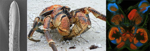
Birgus latro the giant robber crab or coconut crab (Anomura, Coenobitidae) is the largest land-living arthropod in the world and inhabits Indo-Pacific islands such as the Christmas Island. Several previous studies (e.g. Stensmyr et al., 2005) have provided evidence that B. latro has a fine and differentiated sense of smell. The conquest of land occurred probably five times convergently within Crustacea. In this regard, we are interested in terrestrial adaptations of the sensory organs and the nervous system of different crustacean taxa in comparison to their nearest marine relatives. Our focus is on the morphology of central olfactory processing areas. We examined histological cross sections of the brains of B. latro and three other species among the Brachyura and Anomura with immunohistochemistry against synaptic proteins and allatostatin. For these experiments, we analyzed the marine Carcinus maenas (Brachyura), Pagurus bernhardus (Anomura) and the terrestrial Gecarcoidea natalis (Brachyura). In addition, we reconstructed a three dimensional model of the brain and selected brain structures of B. latro. Our aim is to improve the knowledge of the brain and the central olfactory pathway of Crustaceans as well as a better understanding of the different adaptation strategies for conquering land within the Meiura.

Description of a new Spadella-Species (Chaetognatha) in a north adriatic bay
Diploma thesis by Dipl.-Biol. Charlotte Winkelmann
The genus Spadella belongs to the Chaetognatha, a group of marine animals which are adapted to a benthic way and can be found in all seas. The newly described Spadella valsalinae sp. nov. was discovered and collected in the Northern Adriatic Sea in 10–11 m. It is closely related to Spadella cephaloptera, Spadella ledoyeri, Spadella birostrata and compared with the two interstitial species Spadella interstitialis and Spadella nunezi. In order to understand the distribution of the Spadellids, their biogeography in the Mediterranean Sea was studied with the focus on troglophilic species in the western Mediterranean Sea with a coexistence of infralitoral species in an eperic sea as the Adriatic Sea.

